
Skin structure and function
Skin has three layers:
- The epidermis, the outermost layer of skin, provides a waterproof barrier and creates our skin tone.
- The dermis, beneath the epidermis, contains tough connective tissue, hair follicles, and sweat glands.
- The deeper subcutaneous tissue (hypodermis) is made of fat and connective tissue.

Epidermis
The epidermis is stratified squamous epithelium. The main cells of the epidermis are the keratinocytes, which synthesise the protein keratin. Protein bridges called desmosomes connect the keratinocytes, which are in a constant state of transition from the deeper layers to the superficial . The four separate layers of the epidermis are formed by the differing stages of keratin maturation. The epidermis varies in thickness from 0.05 mm on the eyelids to 0.8±1.5 mm on the soles of the feet and palms of the hand.
Moving from the lower layers upwards to the surface, the four layers of the epidermis are:
- stratum basale (basal or germinativum cell layer)
- stratum spinosum (spinous or prickle cell layer)
- stratum granulosum (granular cell layer)
- stratum corneum (horny layer).
In addition, the stratum lucidum is a thin layer of translucent cellsseen in thick epidermis. It represents a transition from the stratum granulosum and stratum corneum and is not usually seen in thin epidermis. Together, the stratum spinosum and stratum granulosum are sometimes referred to as the Malphigian layer.
Dermis
The dermis varies in thickness, ranging from 0.6 mm on the eyelids to 3 mm on the back, palms and soles. It is found below the epidermis and is composed of a tough, supportive cell matrix.
Two layers comprise the dermis:
- a thin papillary layer
- a thicker reticular layer.
The papillary dermis lies below and connects with the epidermis. It contains thin loosely arranged collagen fibres. Thicker bundles of collagen run parallel to the skin surface in the deeper reticular layer, which extends from the base of the papillary layer to the subcutis tissue. The dermis is made up of fibroblasts, which produce collagen, elastin and structural proteoglycans, together with immunocompetent mast cells and macrophages. Collagen fibres make up 70% of the dermis, giving it strength and toughness. Elastin maintains normal elasticity and flexibility while proteoglycans provide viscosity and hydration. Embedded within the fibrous tissue of the dermis are the dermal vasculature, lymphatics, nervous cells and fibres, sweat glands, hair roots and small quantities of striated muscle.
Subcutis
This is made up of loose connective tissue and fat, which can be up to 3 cm thick on the abdomen.

Derivative structures of the skin
Sebaceous glands
These glands are derived from epidermal cells and are closely associated with hair follicles especially those of the scalp, face, chest and back; they are not found in hairless areas. They are small in children, enlarging and becoming active at puberty, being sensitive to androgens. They produce an oily sebum by holocrine secretion in which the cells break down and release theirlipid cytoplasm. The full function of sebum is unknown at present but it does play a role in the following:
- maintaining the epidermal permeability barrier, structure and differentiation
- skin-specific hormonal signalling
- transporting antioxidants to the skin surface
- protection from UV radiation.
The eccrine sweat gland, which is controlled by the sympathetic nervous system, regulates body temperature. When internal temperature rises, the eccrine glands secrete water to the skin surface, where heat is removed by evaporation. If eccrine glands are active over most of the body (as in horses, bears, and humans), they are major thermoregulatory devices. In other animals (dogs, cats, cattle, and sheep), they are active only on the pads of the paws or along the lip margins and may be entirely absent over the rest of the body; such animals often depend on panting for effective temperature control. Smaller mammals, such as rodents, cannot endure dehydration and hence possess no eccrine glands at all.
Apocrine sweat glands, which are usually associated with hair follicles, continuously secrete a fatty sweat into the gland tubule. Emotional stress causes the tubule wall to contract, expelling the fatty secretion to the skin, where local bacteria break it down into odorous fatty acids. In human beings, apocrine glands are concentrated in the underarm and in genital regions; the glands are inactive until they are stimulated by hormonal changes in puberty. In other mammals, apocrine glands are more numerous. Certain specialized glands, such as mammary glands, wax-secreting glands of the ear canal, and many mammalian scent glands, probably developed from modified apocrine glands.
Functions of the skin
- Provides a protective barrier against mechanical, thermal and physical injury and noxious agents.
- Prevents loss of moisture.
- Reduces the harmful efects of UV radiation.
- Acts as a sensory organ.
- Helps regulate temperature control.
- Plays a role in immunological surveillance.
- Synthesises vitamin D3.
- Has cosmetic, social and sexual associations
As the viable cells move towards the stratum corneum they begin to clump proteins into granules in the granular layer. The granules are filled with the protein fillagrin, which becomes complexed with keratin to prevent the breakdown of fillagrin by proteolytic enzymes. As the degenerating cells move towards the outer layer, enzymes break down the keratin-fillagrin complex. Fillagrin forms on the outside of the corneocytes while the waterretaining keratin remains inside. When the moisture content of the skin reduces, fillagrin is further broken down into free amino acids by specific proteolytic enzymes in the stratum corneum. The breakdown of fillagrin only occurs when the skin is dry in order to control the osmotic pressure and the amount of water it holds: in healthy skin the water content of the stratum corneum is normally around 30%. The free amino acids, along with other components such as lactic acid, urea and salts, are known as `natural moisturising factors' (NMF) and are responsible for keeping the skin moist and pliable due to their ability to attract and hold water.
Lipids
The major factor in the maintenance of a moist, pliable skin barrier is the presence of intercellular lipids. These form stacked bilayers that surround the corneocytes and incorporate water into the stratum corneum. The lipids are derived from lamellar granules, which are released into extracellular spaces of degrading cells in the granular layer; the membranes of these cells also release lipids. Lipids include cholesterol, free fatty acids and sphingolipids. Ceramide, a type of sphingolipid, is mainly responsible for generating the stacked lipid structures that trap water molecules in their hydrophilic region. These stacked lipids surround the corneocytes and provide an impermeable barrier by preventing the movement of water and NMF out of the surface layers of the skin. After the age of 40 there is a sharp decline in skin lipids thus increasing our susceptibility to dry skin conditions.
Shedding (desquamation) of skin cells
Shedding the cells of the stratum corneum is an important factor in maintaining skin integrity and smoothness. Desquamation involves the enzymatic process of dissolving the protein bridges, the desmosomes, between the corneocytes, and the eventual shedding of these cells. The proteolytic enzymes responsible for desquamation are located intracellularly and function in the presence of a well-hydrated stratum corneum. In the absence of water the cells do not desquamate normally and the skin becomes roughened, dry, thickened and scaly. In normal healthy skin there is a balance in the production and shedding of corneocytes. In diseases such as psoriasis, which involve increased corneocyte production, and where decreases in shedding occur, the result is dry, rough skin as the cells accumulate on the skin.
Remaining functions
UV protection Melanocytes, located in the basal layer, and melanin have important roles in the skin's barrier function by preventing damage by UV radiation. In the inner layers of the epidermis, melanin granules form a protective shield over the nuclei of the keratinocytes; in the outer layers, they are more evenly distributed. Melanin absorbs UV radiation, thus protecting the cell's nuclei from DNA (deoxyribonucleic acid) damage. UV radiation induces keratinocyte proliferation, leading to thickening of the epidermis.
Thermoregulation
The skin plays an important role in maintaining a constant body temperature through changesin blood flow in the cutaneous vascularsystem and evaporation of sweat from the surface.
Immunological surveillance
As well acting as a physical barrier, skin also plays an important immunological role. It normally contains all the elements of cellular immunity, with the exception of B cells.
Skin Conditions

- Rash: Nearly any change in the skin’s appearance can be called a rash. Most rashes are from simple skin irritation; others result from medical conditions.
- Dermatitis: A general term for inflammation of the skin. Atopic dermatitis (a type of eczema) is the most common form.
- Eczema: Skin inflammation (dermatitis) causing an itchy rash. Most often, it’s due to an overactive immune system.
- Psoriasis: An autoimmune condition that can cause a variety of skin rashes. Silver, scaly plaques on the skin are the most common form.
- Dandruff: A scaly condition of the scalp may be caused by seborrheic dermatitis, psoriasis, or eczema.
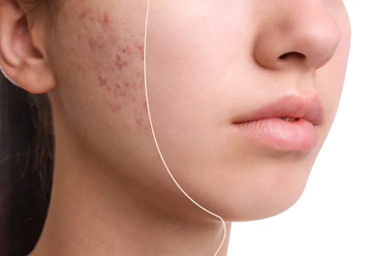
- Acne: The most common skin condition, acne affects over 85% of people at some time in life.
- Cellulitis: Inflammation of the dermis and subcutaneous tissues, usually due to an infection. A red, warm, often painful skin rash generally results.
- Skin abscess (boil or furuncle): A localized skin infection creates a collection of pus under the skin. Some abscesses must be opened and drained by a doctor in order to be cured.
- Rosacea: A chronic skin condition causing a red rash on the face. Rosacea may look like acne, and is poorly understood.
- Warts: A virus infects the skin and causes the skin to grow excessively, creating a wart. Warts may be treated at home with chemicals, duct tape, or freezing, or removed by a physician.
- Melanoma: The most dangerous type of skin cancer, melanoma results from sun damage and other causes. A skin biopsy can identify melanoma.
- Basal cell carcinoma: The most common type of skin cancer. Basal cell carcinoma is less dangerous than melanoma because it grows and spreads more slowly.
- Seborrheic keratosis: A benign, often itchy growth that appears like a “stuck-on” wart. Seborrheic keratoses may be removed by a physician, if bothersome.
- Actinic keratosis: A crusty or scaly bump that forms on sun-exposed skin. Actinic keratoses can sometimes progress to cancer.
- Squamous cell carcinoma: A common form of skin cancer, squamous cell carcinoma may begin as an ulcer that won’t heal, or an abnormal growth. It usually develops in sun-exposed areas.
- Herpes: The herpes viruses HSV-1 and HSV-2 can cause periodic blisters or skin irritation around the lips or the genitals.
- Hives: Raised, red, itchy patches on the skin that arise suddenly. Hives usually result from an allergic reaction.
- Tinea versicolor: A benign fungal skin infection creates pale areas of low pigmentation on the skin.
- Viral exantham: Many viral infections can cause a red rash affecting large areas of the skin. This is especially common in children.
- Shingles (herpes zoster): Caused by the chickenpox virus, shingles is a painful rash on one side of the body. A new adult vaccine can prevent shingles in most people.
- Scabies: Tiny mites that burrow into the skin cause scabies. An intensely itchy rash in the webs of fingers, wrists, elbows, and buttocks is typical of scabies.
- Ringworm: A fungal skin infection (also called tinea). The characteristic rings it creates are not due to worms.
The Effects of Aging on Skin
As is clearly evident from cosmetic advertisements, most of us use the skin as an indicator of aging. Intrinsic aging is manifest in skin areas protected from the sun, such as the buttocks. The additional damage caused by long-term exposure to the sun’s ultraviolet radiation is called photoaging.
In intrinsic aging, the thickness of the epidermis decreases slightly, with no change in the outermost epidermal layer, the stratum corneum. The rate of generation of keratinocytes, which end their lives as the stratum corneum, slows with age, which increases the dwell time of stratum-corneum components. The decreasing number of melanocytes reduces photoprotection, and the decreasing number of Langerhans cells reduces immune surveillance.
Intrinsic aging of the dermis affects mainly the extracellular matrix. The amount of elastin and collagen decreases, and their structures change. Glycosaminoglycan composition also changes. The levels of hyaluronic acid and the proteoglycan versican—which contains chondroitin sulfate (CS) and dermatan sulfate (DS)—are decreased. On the other hand, the level of the proteoglycan decorin—which also contains CS and DS—is increased. As a result, the dermis thins by ~20% and becomes stiffer, less malleable, and thus more vulnerable to injury.
Photoaging increases the extent of most intrinsic age changes in both the epidermis and dermis, and it has additional effects. For example, photoaging causes coarse wrinkles, which occur minimally or not at all because of intrinsic aging.
Aging also reduces the number and function of sweat glands as well as the production of sebum by sebaceous glands.
Why skin care is so important?
Proper skin care is important because our skin is the largest barrier against infection that we have. Keeping our skin healthy and moist helps keep this barrier strong. When the skin gets dry or irritated by harsh soaps, cracks in the skin can occur. Cracks in the skin make a person more prone to infection.
Ozonated oil are wonderful for skin because they...
- Remove the dead layer of cells on your skin’s surface, revealing the younger cells beneath, and leaving your skin feeling soft and smooth.
- Contain natural oils that moisturise and feed your skin.
- Contain ozonide that has a remarkable ability to neutralize toxins, calm inflammation, activate epidermal wound repair, minimize the appearance of pores and age spots, and dramatically improves skin texture.
References
Medical Physiology
WALTER F. BORON, MD, PhD
EMILE L. BOULPAEP, MD
Dermatology
-
J. G. Rycroft, S. J. Robertson, S. H. Wakelin


is chloroquine phosphate the same as hydroxychloroquine hydrochlorothiazide 12.5 mg chloroquin side effects
hydroclorquin resochin why is hydroxychloroquine
side effects tadalafil where to get tadalafil tadalafil otc
tadalafil online with out prescription purchase peptides tadalafil tadalafil cost walmart
tadalafil brands cialis pills tadalafil sublingual